Introduction
Unilateral Biportal Endoscopy (UBE) has emerged as one of the most advanced minimally invasive techniques in modern spine surgery. By using two small portals-one for endoscopic visualization and one for surgical instruments-UBE enables surgeons to perform precise decompression and discectomy with minimal damage to muscles and soft tissues. As a safe, effective, and patient-friendly procedure, UBE is rapidly becoming the preferred choice for various lumbar degenerative diseases worldwide.
Clinical Applications
UBE is widely used to treat common lumbar conditions:
- Lumbar disc herniation (including migrated and sequestered hernias)
- Lumbar spinal stenosis (central, lateral recess and foraminal stenosis)
- Grade I–II degenerative lumbar spondylolisthesis
- Facet joint cysts
- Revision spine surgery
Surgical Process of UBE (Step-by-Step)
1. Patient Positioning & Anesthesia
- Patient lies prone under general or epidural anesthesia.
- C-arm fluoroscopy is used for real-time imaging and level confirmation.
2. Portal Placement (Key Step)
- Two 1.0–1.5 cm skin incisions are made at the target level under fluoroscopic guidance.
- Cranial portal: for endoscope (viewing).
- Caudal portal: for instruments (working).
- Portals are usually 1.5–2 cm apart, over the lamina and facet joint.
3. Soft Tissue Dissection & Exposure
- Serial dilators are inserted to create a clear working space.
- Radiofrequency (RF) ablation removes soft tissue over the lamina, exposing the facet joint, lamina, and ligamentum flavum (LF).
- Continuous saline irrigation maintains a clear surgical field and controls bleeding.
4. Laminectomy & Ligamentum Flavum Resection
- Under endoscopic view, a high-speed burr and Kerrison punch are used to remove part of the ipsilateral lamina (partial hemilaminectomy).
- The ligamentum flavum is carefully dissected and removed to expose the spinal canal and nerve roots.
- For bilateral decompression, the "Z-technique" is used: remove ipsilateral lamina → base of spinous process → contralateral lamina → resect contralateral LF.
5. Nerve Root Decompression & Discectomy
- The nerve root is identified and protected.
- Herniated disc material is removed using pituitary forceps and curettes.
- Osteophytes and hypertrophied facet tissue are trimmed to widen the spinal canal and foramina.
- Complete nerve root decompression is confirmed by free mobilization of the nerve root.
6. Hemostasis & Closure
- RF coagulation is used to achieve hemostasis.
- Irrigation is stopped, and instruments are removed.
- Small incisions are closed with sutures or surgical adhesive.
Clinical Outcomes
Clinical studies show significant improvements after UBE surgery:
- VAS scores: Leg pain from 7.8 → 1.7; back pain from 5.8 → 1.7.
- ODI: From 63.0 → 20.7 (marked reduction in disability).
- Patient satisfaction: 87.3% report good to excellent outcomes (Macnab criteria).
- Complication rate: 5.5%–13.8% (dural tear is most common, usually minor).
Conclusion
UBE represents a major leap forward in minimally invasive spine surgery. It combines the benefits of open surgery's visualization with endoscopy's minimal trauma, delivering safe, effective, and durable results for patients with lumbar degenerative diseases.
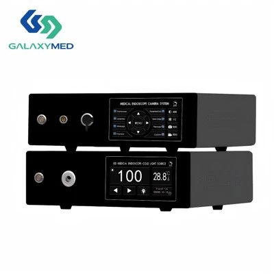
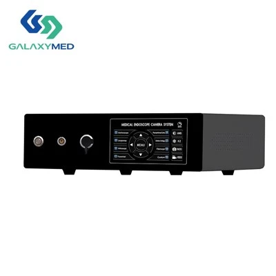
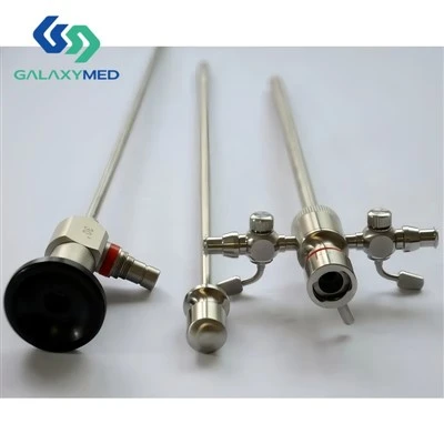
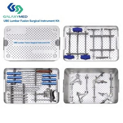
As a professional medical device supplier, we provide full-range UBE instruments and endoscopy systems to support surgeons in performing precise and efficient UBE procedures.